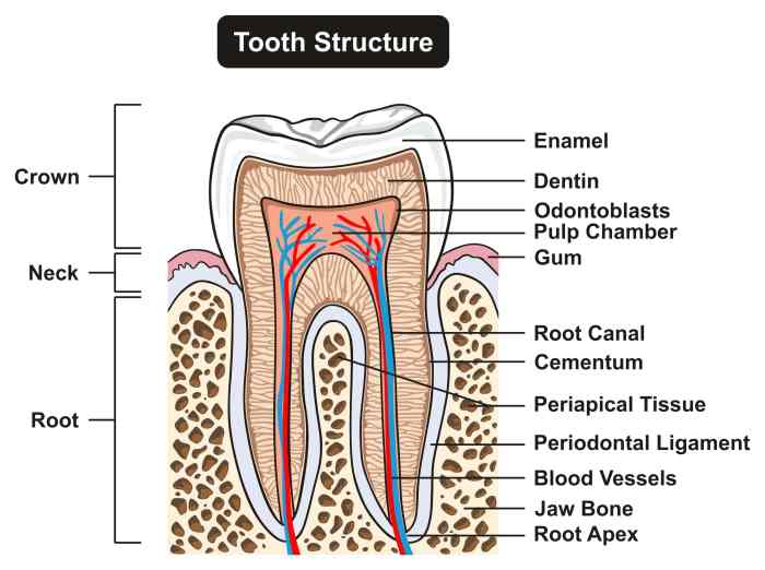
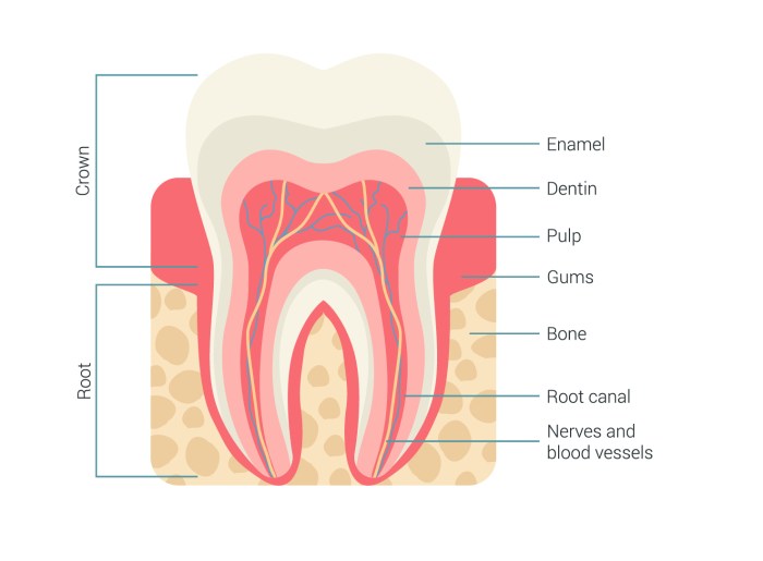
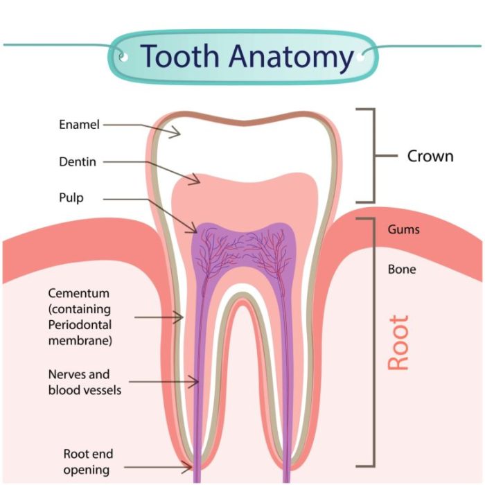
Label the anatomical features of a tooth – Labeling the anatomical features of a tooth is crucial for understanding its structure and function. This guide provides a detailed overview of the crown, root, pulp cavity, dentin, cementum, and periodontal ligament, empowering readers with a comprehensive understanding of this essential oral component.
Crown

The crown is the visible portion of the tooth that extends above the gum line. It has a complex shape that varies depending on the type of tooth. In general, the crown is wider at the base and tapers towards the tip.
The chewing surface of the crown is covered with cusps, which are raised areas that help to grind food.
- Incisors:Have a single, sharp cusp for cutting food.
- Canines:Have a single, pointed cusp for tearing food.
- Premolars:Have two or more cusps for grinding food.
- Molars:Have multiple cusps for grinding food.
The crown is made up of a hard, white substance called enamel. Enamel is the hardest substance in the human body and it helps to protect the crown from decay.
Root

The root is the portion of the tooth that is embedded in the jawbone. It is typically longer than the crown and has a tapered shape. The root is covered with a layer of cementum, which is a hard, calcified tissue that helps to anchor the tooth in the jawbone.
The root also contains the pulp cavity, which is a hollow space that contains the blood vessels, nerves, and connective tissue that nourish the tooth.The root is essential for anchoring the tooth in the jawbone and for providing it with nutrients.
Without a healthy root, the tooth would not be able to function properly.
Pulp Cavity
The pulp cavity is a hollow space that is located in the center of the tooth. It contains the blood vessels, nerves, and connective tissue that nourish the tooth. The pulp cavity is lined with a thin layer of dentin, which is a hard, calcified tissue that helps to protect the pulp.The
pulp cavity is important for maintaining the health of the tooth. If the pulp cavity becomes infected, it can lead to pain, swelling, and even tooth loss.
Dentin
Dentin is a hard, calcified tissue that makes up the bulk of the tooth. It is located beneath the enamel and surrounds the pulp cavity. Dentin is less hard than enamel, but it is still strong enough to protect the pulp cavity from damage.Dentin
is formed by the odontoblasts, which are cells that are located in the pulp cavity. Odontoblasts secrete a matrix of collagen and other proteins, which is then mineralized to form dentin.Dentin is important for supporting the enamel and protecting the pulp cavity.
It also helps to transmit forces from the chewing surface of the tooth to the root.
Cementum: Label The Anatomical Features Of A Tooth

Cementum is a hard, calcified tissue that covers the root of the tooth. It is less hard than enamel or dentin, but it is still strong enough to protect the root from damage. Cementum is formed by the cementoblasts, which are cells that are located in the periodontal ligament.Cementum
is important for attaching the tooth to the periodontal ligament. It also helps to protect the root from damage and to repair any damage that does occur.
Periodontal Ligament
The periodontal ligament is a thin layer of connective tissue that surrounds the root of the tooth. It attaches the tooth to the jawbone and helps to transmit forces from the chewing surface of the tooth to the root. The periodontal ligament also contains blood vessels and nerves that nourish the tooth.The
periodontal ligament is important for supporting the tooth and for transmitting forces during chewing. It also helps to protect the root from damage and to repair any damage that does occur.
Essential Questionnaire
What is the function of the enamel?
Enamel protects the crown of the tooth from wear and tear.
What is the role of the periodontal ligament?
The periodontal ligament supports the tooth and transmits forces during chewing.
How does dentin contribute to tooth structure?
Dentin provides support for the enamel and protects the pulp.